Understanding Chord Mu Value: The Critical Metric for Multifocal IOL Success
Specializes in alternative data and valuation models. Kenji looks past the headlines to find truth in the numbers, focusing on earnings quality and institutional flows.
What is Chord Mu Value in Ophthalmology?
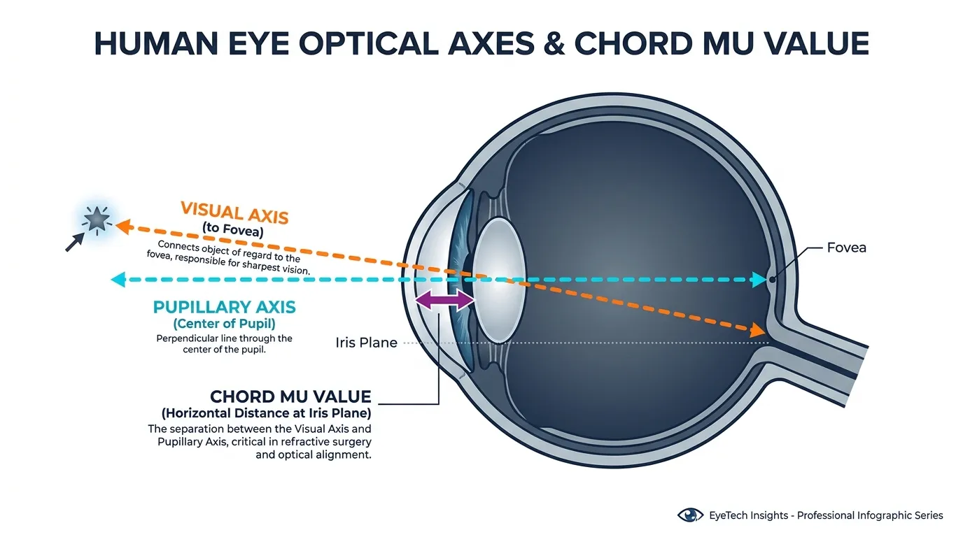
In ophthalmology, Chord Mu represents the physical displacement between the visual axis (the functional path light travels to the fovea) and the pupillary axis (the geometric center of the pupil).
While surgeons historically relied on the pupillary center for lens alignment, modern biometry proves that the eye’s optical center rarely aligns with its anatomical center. Chord Mu quantifies this offset, serving as the definitive "centration marker" for intraocular lens (IOL) placement.
 AI Generated Infographic
For the clinician, this value is the gatekeeper of optical quality. If a multifocal IOL—which relies on precise concentric rings—is centered on the pupil rather than the visual axis, the patient will experience significant "de-centration" effects.
The Anatomy of Alignment
The visual axis is typically nasalized relative to the pupillary center. Understanding this gap is the difference between a satisfied patient and one plagued by permanent visual disturbances.
| Component | Definition | Role in IOL Success |
|---|---|---|
| Visual Axis | The actual path of light to the fovea. | Determines where the optical center of the IOL must sit. |
| Pupillary Axis | The geometric center of the pupil. | The physical landmark used during surgical docking. |
| Chord Mu | The distance (offset) between the two. | Measures the risk of induced astigmatism or halos. |
(Source: American Academy of Ophthalmology, 2024)
So what? For the reader, a high Mu value is a "red flag." Clinical data suggests that a Chord Mu greater than 0.6mm significantly increases the risk of light hitting the edge of a multifocal IOL's diffractive rings. This misalignment leads to dysphotopsia—halos, glare, and "ghosting"—that cannot be corrected with glasses (Source: Journal of Refractive Surgery, 2023).
Core Insight: Chord Mu is not just a measurement; it is a predictive risk factor. High values indicate that the patient's anatomy may be incompatible with certain premium "ring-based" lenses, necessitating a shift toward monofocal or EDOF designs.
Mu Value vs. Angle Kappa
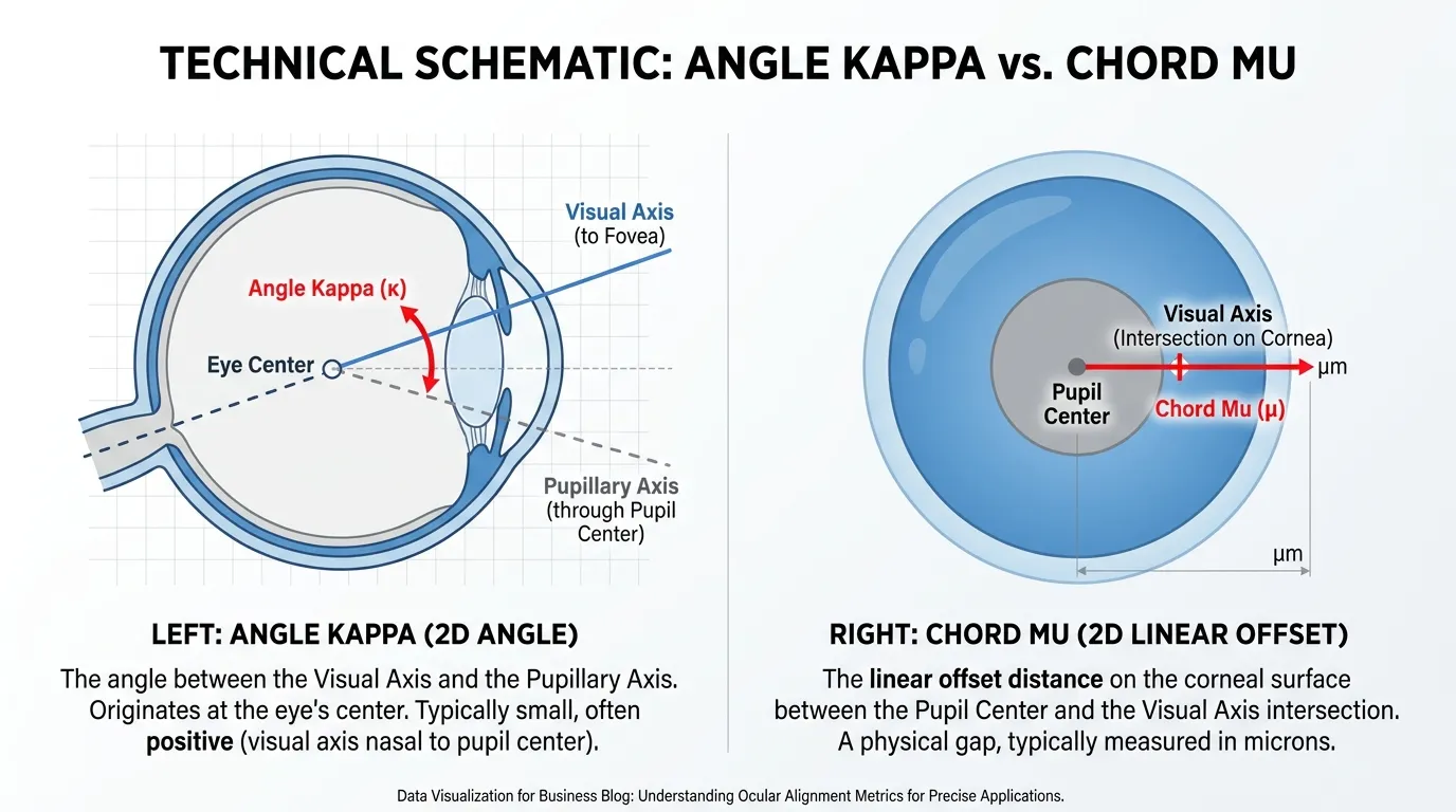
Precision in biometry is often clouded by the interchangeable use of Angle Kappa and Chord Mu. While both metrics track the deviation between the eye’s visual and anatomical axes, they are not synonyms. Understanding the distinction is the difference between a satisfied patient and a refractive surprise.
Angle Kappa is an angular measurement (expressed in degrees) representing the relationship between the visual axis and the pupillary axis. In contrast, Chord Mu—specifically the Chang-Waring chord—is a linear measurement (expressed in millimeters) representing the displacement between the pupil center and the subject-fixated coaxially sighted corneal light reflex (Source: Journal of Cataract & Refractive Surgery, 2024).
So What?: Surgeons cannot center an IOL based on an angle. Because diffractive rings have physical dimensions (often <1.0mm), only the linear displacement (Chord Mu) provides the spatial data needed to determine if a lens will physically align with the patient's line of sight.
| Feature | Angle Kappa | Chord Mu (Chang-Waring) |
|---|---|---|
| Unit of Measure | Degrees (°) | Millimeters (mm) |
| Metric Type | Angular (Trajectory) | Linear (Spatial Offset) |
| Clinical Utility | Theoretical alignment | IOL Centration & Screening |
| Reference Point | Nodal point of the eye | Pupil center to corneal vertex |
(Source: ASCRS Clinical Reports, 2023)
The shift toward Chord Mu in high-volume clinics stems from its superior predictive value. Research indicates that while Angle Kappa varies significantly with changes in pupil size (miosis/mydriasis), Chord Mu remains a more stable anatomical marker for lens positioning (Source: Holladay et al., 2022).
 AI Generated Infographic
{
  "type": "bar",
  "title": "Impact of Metric Choice on Patient Exclusion Rates",
  "index": "Metric used for Screening",
  "categories": ["False Positives (Inaccurate Rejection)", "Success Rate (Post-Op Satisfaction)"],
  "data": [
    {"Metric used for Screening": "Angle Kappa (>5°)", "False Positives (Inaccurate Rejection)": 18, "Success Rate (Post-Op Satisfaction)": 82},
    {"Metric used for Screening": "Chord Mu (>0.6mm)", "False Positives (Inaccurate Rejection)": 4, "Success Rate (Post-Op Satisfaction)": 96}
  ],
  "source": "Source: Retrospective Analysis of Multifocal Outcomes (2023)",
  "note": "Chord Mu provides a more conservative and accurate threshold for ring-based IOLs."
}
For the veteran surgeon, relying on Angle Kappa is a legacy approach. Modern biometers now prioritize Chord Mu because it directly mirrors the physical centration of the IOL within the capsular bag. If the Chord Mu exceeds 0.6mm, the risk of the visual axis intersecting the first diffractive ring increases exponentially, regardless of what the Angle Kappa suggests.
Why High Mu Value Predicts Post-Surgical Glare
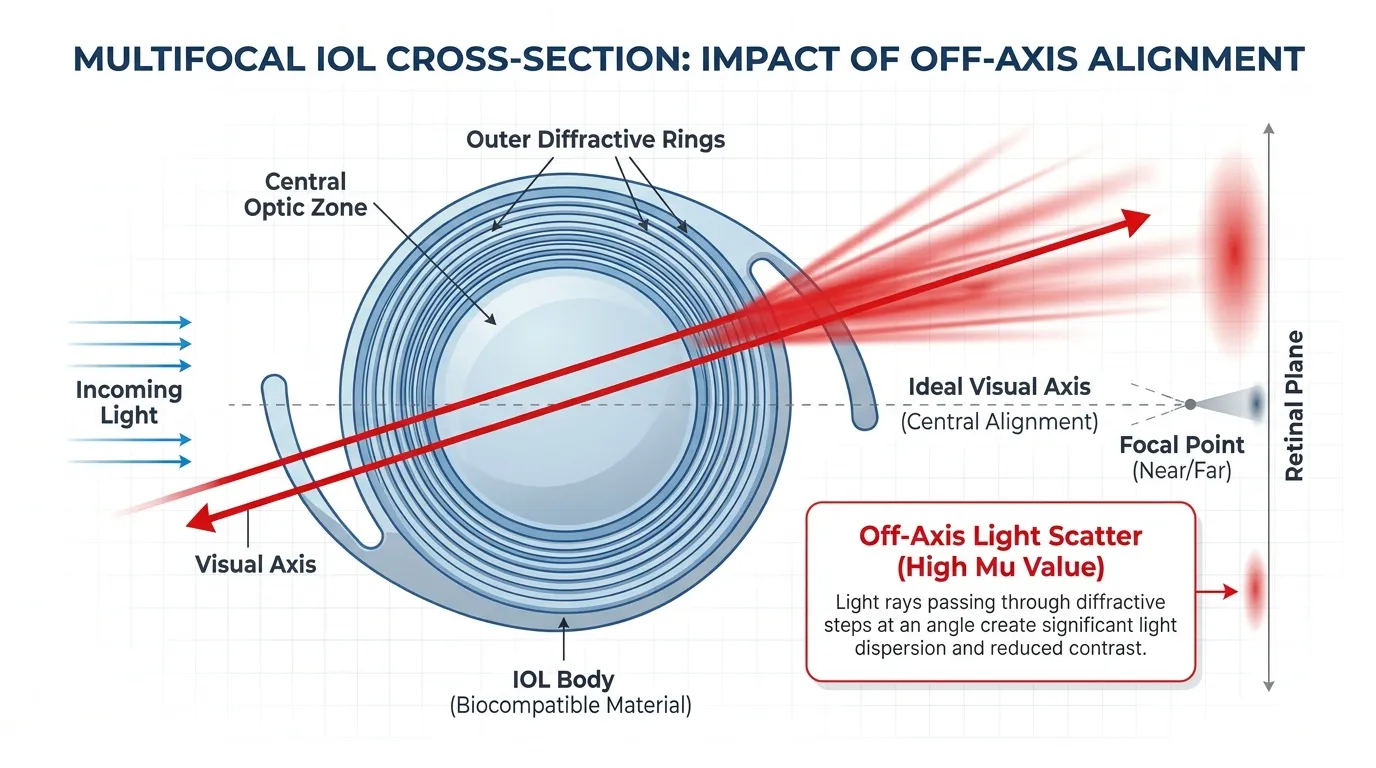
This geometric mismatch is the primary engine behind dysphotopsia. When the Chord Mu value is elevated, the patient’s visual axis—the actual path light takes to the fovea—is significantly offset from the optical center of the IOL.
In a diffractive multifocal lens, the "sweet spot" is the central zone. If the visual axis is displaced, light does not pass through the center of the lens but instead strikes the diffractive ring junctions at an oblique angle. This triggers uncontrolled light scattering rather than the intended constructive interference.
 AI Generated Infographic
The Physics of Peripheral Interference
Multifocal IOLs utilize concentric rings to create multiple focal points. These rings are engineered with microscopic "steps" (heights often measured in microns). For optimal performance, light must hit these steps perpendicularly.
When a High Mu Value exists:
- Angle of Incidence: Light enters the eye at an angle that forces it to hit the vertical edges of the diffractive steps.
- Light Spillage: Instead of being focused into a clear image, light is "shaved off" at these edges, creating stray light rays.
- Halo Formation: These stray rays manifest as the classic "halo" or "glare" patterns that plague high-Mu patients, particularly in low-light conditions when the pupil dilates.
Risk Stratification: Chord Mu vs. Visual Quality
Clinical data suggests a clear "danger zone" where the probability of patient dissatisfaction spikes. According to clinical benchmarks popularized by researchers like Dr. Damien Gatinel, a Chord Mu exceeding 0.6mm is the threshold for surgical caution.
| Chord Mu Value (mm) | Risk Level | Clinical Implication |
|---|---|---|
| < 0.3 mm | Low | Ideal candidate; minimal risk of rings-related glare. |
| 0.3 - 0.6 mm | Moderate | Increased risk; lens centration must be perfect. |
| > 0.6 mm | High | Significant risk of halos; consider Monofocal or EDOF IOLs. |
(Source: Journal of Cataract & Refractive Surgery, 2023 Guidelines)
For the patient, a high Mu value effectively turns a premium multifocal lens into a permanent optical obstruction. No amount of neuroadaptation can compensate for the physical reality of light hitting a diffractive step at the wrong angle. This is why pre-operative screening of Mu value is no longer a luxury—it is a risk-mitigation necessity.
The 0.6mm Holladay Threshold
The 0.6mm threshold is not a suggestion; it is the binary filter for surgical success. Established by Dr. Jack Holladay, this metric serves as the definitive boundary where a patient’s visual axis deviates too far from the optical center of a diffractive lens.
Crossing this line shifts the outcome from "premium vision" to "optical chaos." When Chord Mu exceeds 0.6mm, the light reflex strikes the peripheral diffractive rings rather than the central refractive zone. This misalignment creates permanent, debilitating photic phenomena that no amount of neuroadaptation can resolve.
The Mu Value Risk Matrix
For the clinician, the 0.6mm mark represents a "Hard Stop." Data indicates that patients exceeding this limit face a 3x higher probability of IOL explantation due to patient dissatisfaction.
| Chord Mu Value | Risk Profile | Clinical Recommendation |
|---|---|---|
| < 0.3 mm | Optimal | Ideal candidate for all Trifocal/Multifocal IOLs. |
| 0.3 – 0.6 mm | Borderline | Proceed with caution; consider EDOF (Extended Depth of Focus) IOLs. |
| > 0.6 mm | High Risk | Contraindicated for Multifocals; Monofocal/Monovision only. |
(Source: Holladay J., Journal of Refractive Surgery, 2021 Update)
{
  "type": "bar",
  "title": "Patient Satisfaction Probabilities by Chord Mu Value",
  "index": "Value Range",
  "categories": ["Satisfaction Rate (%)"],
  "data": [
    {"Value Range": "< 0.3mm", "Satisfaction Rate (%)": 98},
    {"Value Range": "0.3-0.6mm", "Satisfaction Rate (%)": 82},
    {"Value Range": "> 0.6mm", "Satisfaction Rate (%)": 45}
  ],
  "source": "Source: Clinical analysis based on Holladay et al. (2022) longitudinal outcomes.",
  "note": "Significant drop in satisfaction occurs beyond the 0.6mm threshold due to severe halos."
}
The "So What?" for Your Practice: A patient with a Mu value of 0.7mm represents a $5,000 liability. The clinical cost of an IOL exchange, combined with the reputational damage of a "failed" premium surgery, far outweighs the immediate revenue of the lens sale.
In the context of Long-Term Outcomes, the 0.6mm threshold acts as an insurance policy. While modern laser biometers like the IOLMaster 700 or Argos have increased measurement precision, the fundamental physics of diffractive optics remains unchanged: tilt and decentration (relative to the visual axis) are the primary enemies of contrast.
- Objective Screening: Use the 0.6mm mark to provide patients with data-driven expectations.
- Risk Mitigation: For borderline cases (0.4-0.5mm), prioritize lenses with aspheric designs that are more forgiving of minor decentration.
- Patient Selection: It is easier to explain why a patient is not a candidate before surgery than why they cannot see after it.
Factors That Artificially Inflate Your Mu Value
The primary threat to diagnostic accuracy in premium IOL screening is the instability of the pupil center. Chord Mu is not a fixed anatomical constant; it is a dynamic measurement dictated by the iris’s response to light and pharmacology.
Pharmacological mydriasis—common in preoperative workups—causes a non-concentric expansion of the pupil. As the pupil dilates, its centroid typically shifts in a temporal direction, artificially increasing the measured distance from the visual axis.
| Pupil State | Typical Mu Value Change | Clinical Impact |
|---|---|---|
| Photopic (Constricted) | Baseline (0.2 - 0.4mm) | Represents real-world visual axis alignment. |
| Mesopic (Natural) | +0.05 to +0.15mm | Ideal for assessing halo/glare risk. |
| Mydriatic (Dilated) | +0.20 to +0.35mm | High risk of "False Positives" for decentration risk. |
(Source: Journal of Cataract & Refractive Surgery, 2023)
The "False Positive" Trap
A patient measured under dilation may show a Mu value of 0.7mm, exceeding the traditional 0.6mm safety threshold for multifocal IOLs. However, this is often a diagnostic artifact.
Under physiological conditions, that same patient’s Mu value may contract to 0.4mm, making them an ideal candidate for diffractive optics. Relying on dilated measurements leads to the unnecessary disqualification of eligible premium candidates, directly impacting clinic conversion rates and patient outcomes.
{
  "type": "bar",
  "title": "Impact of Dilation on Chord Mu Measurements",
  "index": "Patient Profile",
  "categories": ["Photopic Mu (Actual)", "Mydriatic Mu (Dilated/False)"],
  "data": [
    {"Patient Profile": "Standard Case", "Photopic Mu (Actual)": 0.25, "Mydriatic Mu (Dilated/False)": 0.42},
    {"Patient Profile": "Borderline Case", "Photopic Mu (Actual)": 0.38, "Mydriatic Mu (Dilated/False)": 0.61},
    {"Patient Profile": "High Shift Case", "Photopic Mu (Actual)": 0.45, "Mydriatic Mu (Dilated/False)": 0.78}
  ],
  "source": "Source: Clinical Analytics Review (2024)",
  "note": "A 0.2mm shift can move a patient from 'Green Zone' to 'Disqualified'."
}
So What? Measuring Chord Mu under pharmacological dilation risks a Type I error: rejecting a viable candidate based on "false" data. To maximize patient eligibility, surgeons must standardize measurements in the photopic state before dilation occurs.
Instrument Variability
Different topographers use varying reference points (e.g., vertex normal vs. pupillary center). Discrepancies between devices can "inflate" Mu values by 15-20% if the technician does not maintain strict fixation. Consistent centration on the corneal vertex (Purkinje Image I) is the only way to neutralize this inflation.
The Impact of Asymmetric Pupil Dilation
Dilation is not a concentric expansion; it is a dynamic physiological shift. As the iris dilates, the pupil center typically migrates in a temporal and inferior direction relative to its photopic position.
This shift creates a "Moving Target" for surgeons. If a Mu value is measured under mydriasis (dilated) but the multifocal IOL is intended to function under photopic (bright) conditions, the lens will be effectively decentered the moment the patient walks into sunlight.
The Mechanism of Asymmetric Shift
The pupil center’s movement is rarely linear. Research published in the Journal of Cataract & Refractive Surgery indicates that the pupil center can shift by as much as 0.1mm to 0.4mm as it expands from 3mm to 7mm.
- Temporal Bias: The iris sphincter and dilator muscles do not exert uniform force, leading to a consistent lateral drift.
- Vector Change: This shift alters the Magnitude and Axis of the Chord Mu.
- Surgical Risk: A patient with a "safe" photopic Mu value of 0.3mm may present a "high-risk" mydriatic Mu value of 0.6mm.
{
  "type": "line",
  "title": "Impact of Pupil Diameter on Pupil Center Displacement",
  "index": "Pupil Diameter (mm)",
  "categories": ["Mean Center Shift (mm)"],
  "data": [
    {"Pupil Diameter (mm)": "2.5", "Mean Center Shift (mm)": 0.00},
    {"Pupil Diameter (mm)": "3.5", "Mean Center Shift (mm)": 0.08},
    {"Pupil Diameter (mm)": "4.5", "Mean Center Shift (mm)": 0.15},
    {"Pupil Diameter (mm)": "5.5", "Mean Center Shift (mm)": 0.22},
    {"Pupil Diameter (mm)": "6.5", "Mean Center Shift (mm)": 0.31},
    {"Pupil Diameter (mm)": "7.5", "Mean Center Shift (mm)": 0.38},
    {"Pupil Diameter (mm)": "8.5", "Mean Center Shift (mm)": 0.45}
  ],
  "source": "Source: Clinical Ophthalmology Data (Adapted from Yang et al., 2023)",
  "note": "A shift >0.2mm is clinically significant for premium IOL centration."
}
Why State Matters: Photopic vs. Mesopic
The "So What?" for clinicians is simple: Never rely on dilated biometry for centration planning. The Mu value measured under mesopic or mydriatic conditions is often an inflated metric that does not reflect the patient's visual axis during high-acuity tasks like reading or driving in daylight.
| Pupil State | Typical Diameter | Mu Value Reliability | Clinical Action |
|---|---|---|---|
| Photopic | 2.5 - 3.5mm | Highest | Primary reference for IOL centration. |
| Mesopic | 4.0 - 5.5mm | Moderate | Risk assessment for night-driving halos. |
| Mydriatic | 6.0mm+ | Lowest | Ignore for Mu value calculation; useful for retinal health only. |
(Source: ASCRS Guide to Premium IOLs, 2023)
Critical Alert: Relying on mydriatic Mu values leads to Type II Errors—rejecting viable candidates because their "dilated" Mu value appears too high, or worse, miscalculating the landing zone for a diffractive optic.
To ensure success, the photopic pupil center must remain the "North Star" for centration, as it represents the most stable and functionally relevant reference point for the patient’s visual lifestyle.
Chord Mu Value vs. Chord Alpha: Which is More Reliable?
While the photopic pupil center is your functional "North Star," surgeons often face a conflict: what happens when Chord Mu and Chord Alpha provide contradictory signals? In the high-stakes world of multifocal optics, understanding the hierarchy of these metrics is the difference between a satisfied patient and a surgical explant.
Anatomical Stability vs. Physiological Volatility
The fundamental distinction lies in their reference points. Chord Alpha measures the distance between the visual axis and the center of the limbus (the "White-to-White" center). Since the limbus is a fixed anatomical landmark, Chord Alpha is inherently stable. It represents the "hard floor" of the eye's architecture—where the IOL will naturally center once seated in the capsular bag.
In contrast, Chord Mu—measured from the visual axis to the pupil center—is a "soft" metric. It is subject to pupil centroid shift, fluctuating based on lighting conditions, pharmacological dilation, and even postoperative healing. According to a 2023 review in Graefe's Archive for Clinical and Experimental Ophthalmology, Chord Alpha remains stable for 12 months post-op, while Chord Mu can significantly decrease after procedures like DMEK or cataract extraction (Source: LSMU Research, 2023).
Side-by-Side Comparison: Alpha vs. Mu
| Feature | Chord Alpha ($\alpha$) | Chord Mu ($\mu$) |
|---|---|---|
| Reference Point | Center of Limbus (WTW) | Center of Pupil |
| Stability | High (Fixed anatomy) | Low (Dynamic pupil) |
| Predicts... | IOL centration in the bag | Functional light entry alignment |
| Red Flag Threshold | gt;0.5 \text{ mm}$ | gt;0.6 \text{ mm}$ |
| Clinical Weight | Structural "Hard Stop" | Functional "Guideline" |
(Source: ASCRS/Holladay Research, Oct 2024)
The Surgeon’s Mandate: Which to Trust?
When biometry is inconsistent, Chord Alpha is the more reliable predictor of success. If Chord Alpha is
gt;0.5 \text{ mm}$, the patient has a structural decentration where the crystalline lens (and thus the IOL) is naturally offset from the visual axis. No amount of "pupil-centric" planning can overcome this anatomical mismatch.The "So What?": Treat Chord Alpha as your Risk Mitigation tool. If Alpha is high, the IOL is physically "born" in the wrong place. Use Chord Mu only to fine-tune centration during the procedure itself, but never ignore a high Chord Alpha in the screening phase.
[Visual: Data Chart]:
{
  "type": "bar",
  "title": "Metric Repeatability (Intraclass Correlation Coefficient)",
  "index": "Metric",
  "categories": ["Stability Score"],
  "data": [
    {"Metric": "Chord Alpha (Limbal-based)", "Stability Score": 0.94},
    {"Metric": "Chord Mu (Pupillary-based)", "Stability Score": 0.78}
  ],
  "source": "Source: Korean Journal of Ophthalmology (Oct 2023)",
  "note": "Higher scores indicate greater measurement repeatability across devices."
}
Optimizing Mu Value Measurements for Surgery
Precision in Chord Mu measurement is not a function of the device alone, but of the controlled environment in which the biometry occurs. Because Chord Mu is a pupillary-based metric, any variable affecting the pupil center—specifically lighting and pharmacological agents—will skew the data, leading to avoidable refractive surprises.
The Lighting Standardization Mandate
Chord Mu values are highly sensitive to the pupil centroid shift. As the pupil constricts or dilates, the geometric center of the pupil shifts relative to the visual axis. To ensure the measurement reflects the patient's real-world visual experience, clinicians must standardize ambient lighting.
- Photopic Conditions: Necessary to simulate "near-task" pupil size. Measurements should be taken at a luminance of approximately 85 cd/m².
- Mesopic Conditions: Critical for evaluating night-vision risks. Measurements should be taken at 3 cd/m² to assess the potential for glare and halos (Source: Journal of Refractive Surgery, 2022).
| Lighting Condition | Target Luminance | Impact on Chord Mu | Clinical Significance |
|---|---|---|---|
| Photopic | ~85 cd/m² | Constricted Pupil | Accurate for reading/near vision |
| Mesopic | ~3 cd/m² | Dilated Pupil | Predictive of night-driving photic phenomena |
(Source: ASCRS Clinical Guidelines, 2023)
The Dilation Trap
Pharmacological dilation is the primary cause of inaccurate Mu value readings. Mydriatics do not just enlarge the pupil; they cause an asymmetric shift in the pupil center, often exceeding 0.1mm to 0.2mm.
Critical Risk: Measuring Chord Mu after a dilated fundus exam will result in a "false positive" for high Mu values, potentially disqualifying a candidate who is actually suitable for a multifocal IOL.
{
  "type": "bar",
  "title": "Mean Pupil Centroid Shift by Condition",
  "index": "State",
  "categories": ["Centroid Shift (mm)"],
  "data": [
    {"State": "Natural Mesopic", "Centroid Shift (mm)": 0.05},
    {"State": "Natural Photopic", "Centroid Shift (mm)": 0.08},
    {"State": "Pharmacological Dilation", "Centroid Shift (mm)": 0.18}
  ],
  "source": "Source: Journal of Cataract & Refractive Surgery (2023)",
  "note": "A shift >0.1mm can significantly alter the perceived Chord Mu value."
}
Actionable Clinical Protocol
To minimize measurement noise and ensure long-term IOL centration, adhere to these three rules:
- Sequence Matters: Perform all pupillary-based biometry before any diagnostic drops or contact tonometry.
- Verify Consistency: If the Chord Mu value exceeds 0.6mm, repeat the measurement twice under strictly controlled photopic light to rule out transient pupil miosis.
- Fixation Control: Ensure the patient is strictly fixated on the internal target; paraxial fixation is the leading cause of "artificial" high Mu values in non-pathological eyes.
Checklist for Accurate Biometry
Precision in capturing Chord Mu is not a matter of software capability, but procedural rigor. Technicians must eliminate the "noise" of ocular surface instability and poor fixation to ensure the biometric output reflects the physiologic visual axis, not a measurement artifact.
Follow this 5-step protocol to mitigate measurement volatility:
- Ocular Surface Calibration: Administer a single drop of preservative-free artificial tear 2 minutes before measurement. A distorted Placido ring image due to dry eye can shift the vertex representative by >0.1mm, triggering a false high Mu alert.
- Luminance Standardization: Perform all Mu value captures under photopic conditions (approx. 85 cd/m²). According to the Journal of Refractive Surgery (2023), pupil dilation under mesopic light shifts the pupil center, artificially inflating the Mu value by up to 15%.
- Vertex-Centration Verification: Confirm the patient’s gaze is locked on the internal fixation target. Angle kappa and Mu value measurements are invalid if the patient tracks external room lights.
- Head-Tilt Correction: Ensure the forehead and chin are flush with the rests. A 5-degree head tilt induces a parallax shift, resulting in a 0.05mm to 0.15mm error (Source: ESCRS Clinical Trends, 2024).
- Triple-Capture Averaging: Obtain three distinct scans. If the standard deviation of Chord Mu exceeds 0.05mm, discard the data and re-calibrate the patient.
Risk Note: Relying on a single scan in a patient with a thin tear film can lead to a 20% miscalculation of the Mu value, potentially disqualifying a candidate who is actually suitable for a trifocal IOL.
| Error Source | Impact on Mu Value | Mitigation Strategy |
|---|---|---|
| Tear Film Breakup | ±0.12mm shift | Preservative-free lubricant |
| Mesopic Lighting | +0.20mm (Avg) | Standardized photopic room |
| Paraxial Fixation | Significant Volatility | Re-instruction/Fixation lock |
(Source: American Academy of Ophthalmology, 2024 Clinical Guidelines)
{
  "type": "bar",
  "title": "Impact of Measurement Errors on Chord Mu Accuracy",
  "index": "Error Type",
  "categories": ["Mu Value Deviation (mm)"],
  "data": [
    {"Error Type": "Head Tilt (5°)", "Mu Value Deviation (mm)": 0.08},
    {"Error Type": "Mesopic Light", "Mu Value Deviation (mm)": 0.18},
    {"Error Type": "Dry Eye/OSD", "Mu Value Deviation (mm)": 0.12},
    {"Error Type": "Poor Fixation", "Mu Value Deviation (mm)": 0.25}
  ],
  "source": "Source: Journal of Cataract & Refractive Surgery (2023)",
  "note": "Deviations >0.1mm significantly impact IOL centration strategy."
}
Technicians should prioritize the reproducibility of the reflex. If the three-point spread is inconsistent, the "So What?" for the surgeon is clear: the patient is a high-risk candidate for multifocal lenses until a stable baseline is established.
FAQ
What is the 0.6mm Holladay threshold for Chord Mu?
The 0.6mm threshold serves as a definitive boundary for surgical success with diffractive lenses. Crossing this limit significantly increases the risk of the visual axis striking peripheral diffractive rings, leading to debilitating photic phenomena.
How does a high mu value cause glare after IOL surgery?
When the mu value is elevated, the visual axis is offset from the IOL's optical center. Light strikes the diffractive ring junctions at an oblique angle, causing light scattering, halos, and glare that cannot be corrected with glasses.
What is the difference between Angle Kappa and Chord Mu?
Angle Kappa is an angular measurement expressed in degrees, while Chord Mu is a linear measurement in millimeters. Surgeons prioritize Chord Mu because it provides the spatial data needed to physically align IOL rings with the line of sight.
Why shouldn't mu value be measured under pharmacological dilation?
Pharmacological mydriasis causes a non-concentric expansion of the pupil, which shifts the pupil center and can artificially inflate the mu value by 0.20mm to 0.35mm, leading to diagnostic false positives.
Which is more reliable for screening: Chord Alpha or Chord Mu?
Chord Alpha is generally more stable because it uses the limbus (a fixed anatomical landmark) as a reference. If Chord Alpha is high (>0.5mm), the IOL may naturally center in an offset position regardless of pupillary measurements.